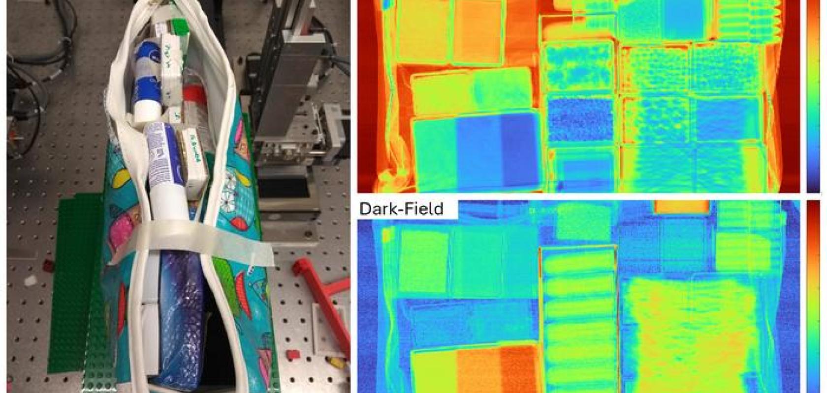
Research conducted at UCL shows that multi-contrast images, generated by combining different x-ray imaging technologies like attenuation and dark field with machine learning, can improve the detection of threatening materials.
The x-ray machines commonly found in airports, as well as those in medical facilities, are based on x-ray attenuation, which reads the x-rays’ reduction in intensity when they pass through different materials. By combining this conventional x-ray attenuation technique’s data with x-ray phase information from refraction and dark-field channels, the new proposed technique creates multi-contrast images to give a better view.
“This method is particularly well suited to discriminating objects with very similar elemental compositions,” said Thomas Partridge from University College London (UCL), who led the research team. “It could be used in airport security or any inline scanning operation to examine materials flagged as suspicious by an initial fast scan such as a traditional x-ray system.”
The team’s research, published in Optica, involved almost 4,000 scans of threatening and non-threatening materials hidden inside bags or obscured by other objects. With a near-perfect recall rate of 99.68% and just one false negative from the threat-baring cases, the new technique was proven to be highly effecting in detecting and identifying explosives.
Improving security with more complex scanners
“Many explosives and common everyday items are composed primarily of carbon, hydrogen, nitrogen and oxygen,” said Partridge, “a similarity that makes them difficult to separate with x-ray attenuation alone. The additional channels offer significantly better enhancement of edges as well as textures and grains of materials, allowing the discrimination of objects with very similar elemental compositions.”
The experiment used to test the new scanning system involved changing the resolution by modifying the scanning speed and applied edge illumination phase contrast. As edge illumination involves placing masks before and after the sample to create the sub-pixel x-ray beamlets needed to make the system sensitive to phase signals, the increased complexity of the system required more sophisticated protocols.
In response to this, the researchers used machine learning with a hierarchical architecture that separates the cluttering objects before distinguishing material types, making it possible to rapidly discern subtle differences in shapes and textures, and distinguish materials based on key identifying features.
Testing the system to accurately find threatening materials
To test the system, a total of 19 threatening and 56 non-threatening materials across three thicknesses were hidden amongst a variety of common non-threatening objects such as brushes, face wipes, socks and other such carry-on items. Using the multi-contrast method in combination with deep learning to analyse the signals, the researchers successfully found 312 threats out of a possible 313, and were not only able to discriminate between materials, but in some cases to identify them, too.
In order to translate the findings to a commercial setting, the researchers are now looking to improve the scanning speed, with one area of active study being to combine the method with 3D computed tomography scanning, which could provide detailed, three-dimensional images of objects. Meanwhile, the findings could have wider implications in other sectors.
“Although more work is needed,” suggested Partridge, “this approach could also prove useful for medical imaging. While traditional x-ray imaging struggles to separate healthy from diseased tissue, other studies have suggested that phase contrast imaging might be able to capture textures that could be used to distinguish healthy and benign tissues.”


