A computer vision-based method is being used to continuously measure mouse body mass non-intrusively, offering a new resource for researchers that promises to enhance the quality of a wide range of pre-clinical studies.
The approach aims to reduce the stress associated with traditional weighing techniques, improving the quality and reproducibility of biomedical research involving mice.
Stressful procedure that can impact clinical results
In both human health and biomedical research, body mass is a critical metric, often serving as an indicator of overall health and a predictor of potential health issues. For researchers working with mice – the most common subjects in preclinical studies – measuring body mass has traditionally involved removing the animals from their cages and placing them on a scale. This process can be stressful for the mice, introducing variables that can affect the outcome of experiments. Moreover, these measurements are typically taken only once every few days, further complicating the accuracy and reproducibility of data.
“We recognised the need for a better method to accurately and noninvasively measure animal mass over time,” said Associate Professor Dr Vivek Kumar, who led the study at The Jackson Laboratory For Genomic Medicine in the US. “The traditional approach not only stresses the mice but also limits the frequency and reliability of measurements, which can weaken the validity of experimental results.”
Computer vision can calculate body mass with <5% error
To address this challenge, Kumar and his team of computational scientists and software engineers, turned to computer vision technology. By analysing one of the largest mouse video datasets used by Kumar previously to also assess grooming behaviour and gait posture, they developed a method to calculate body mass with less than 5% error.
The findings, recently published in Patterns, offer a new resource for researchers that promises to enhance the quality of a wide range of preclinical studies.
Machine vision tools help improve accuracy
The research team faced several challenges in developing this method. Unlike the relatively static subjects used in industrial farming for body mass measurement, mice are highly active and flexible, frequently changing posture and shape, much like a deformable object.
Additionally, the team worked with 62 different mouse strains, ranging in mass from 13 grams to 45 grams, each with unique sizes, behaviours, and coat colours, necessitating the use of multiple visual metrics, machine learning tools, and statistical modelling to achieve the desired level of accuracy.
“In the video data, only 0.6% of the pixels belonged to each mouse, but we were able to apply computer vision methods to predict the body mass of individual mice,” explained first author Malachy Guzman. “By training our models with genetically diverse mouse strains, we ensured that they could handle the variable visual and size distributions commonly seen in laboratory settings. Ultimately our statistical models to predict mass can be used to carry out genetic and pharmacological experiments.”
This new method offers several key advantages for researchers. It enables the detection of small but significant changes in body mass over multiple days, which could be crucial for studies involving drug or genetic manipulations. Additionally, the method has the potential to serve as a diagnostic tool for general health monitoring and can be adapted to different experimental environments and other organisms in the future.
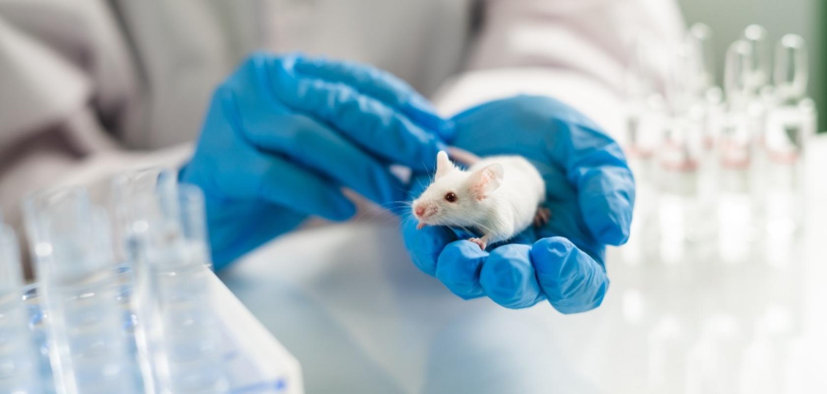
Credit for main image: Egoreichenkov Evgenii/Shutterstock